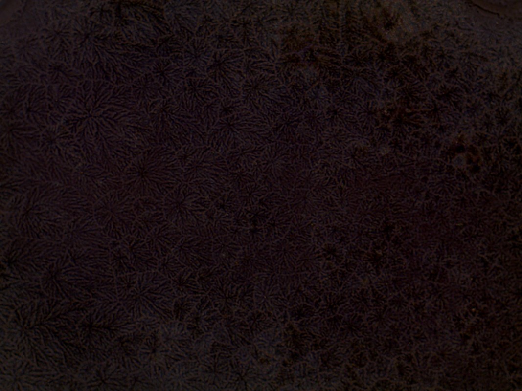
This past week we started feeding our cells using our experimental media. I fed them on Monday and we waited a few days to let the media settle with them. At first our cells seemed to be fine because they looked like they were starting to differentiate and grow. The second day we looked at our cells that had been treated with the Kinky Pink and they did not look good, there were large lumps of what looked like bacteria but turned out to be the sugar crystallizing. When we looked at the cells that were not treated with Kinky Pink they also appeared to have a bacteria like substance growing, luckily it was just on the top of the well plate and we were able to remove it with ethanol.
The final day of our project using the stem cells was spent trypsinizing our cells and dying them with Trypan Blue to do cell counts. We started off by removing the old feeding medium and replacing it with Trypsin to unstick the cells from the bottom of the well plate, we then added our feeding medium back in to stop the Trypsin. After stopping the Trypsin we took some of that mixture and mixed it with the Trypan Blue, we took that new mixture and used the Hemocytometer to take cell counts.
So far we haven't completely looked over our data but from what I've seen in the photos our initial hypothesis doesn't seem to match our data. Unfortunately we are unable to draw a solid conclusion because of the contamination and because we didn't use our experimental media for a long enough period of time to get any real results. If we were to do this project again I would have liked to see how the alcohol really affects the adipocytes after a longer period of time because the initial question we had was left unanswered. We never really got to see if it was the alcohol itself that causes the increase in size or if it was the additives in the alcohol.
The only pattern I really see in our data is that the confluency of our cells hasn't gone beyond 3% and you can see that in the photos of our cells (First one below). There are no real outliers with our data so far everything seems pretty consistent with our cells, the confluency has stayed the same and nothing else has really differed. Our experiment has been completed and our data has been collected. At the end of the experiment I was expecting to see a larger difference between the cells in the ethanol and the ones in the Kinky Pink mixture but they were almost identical in their appearance. Since our experiment has been completed the only additional data we could collect is the size of our cells using the photos we took but with that it would be difficult since there aren't many variations in size. I think right now the only data analysis we can currently do is just look over our final photos and compare them to the photos of the adipocytes before the treatment, the only issue with that is the contamination in the first trial threw us a couple steps back and that data wouldn't really serve a great purpose since they wouldn't match the second trial. We would however be able to take the before photos of the adipocytes from other groups who worked with them and compare them to our final ones.
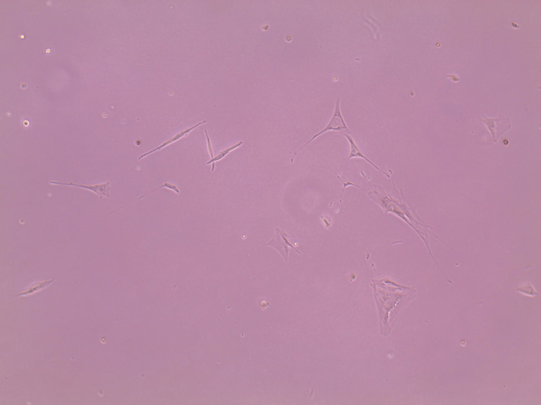
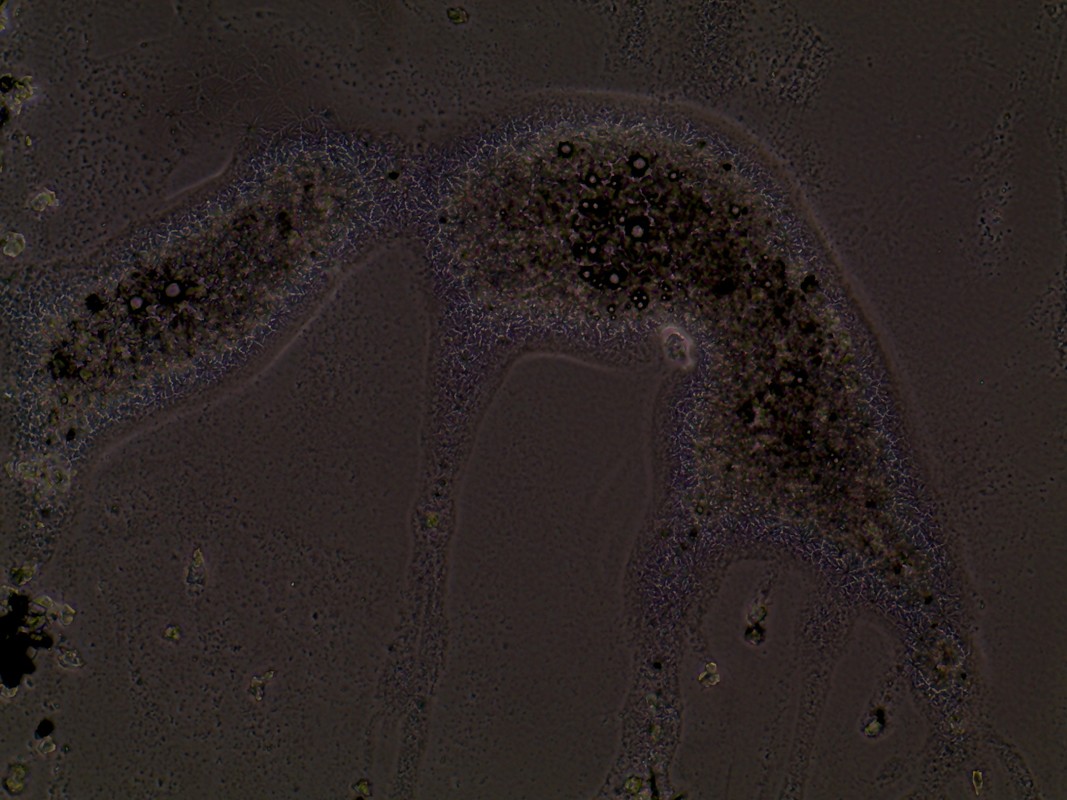
The photos below show our adipocytes in our Kinky Pink mixture (First), the slight contamination we had (Middle) and the crystallized sugar (Last).
Data Sheet
The final day of our project using the stem cells was spent trypsinizing our cells and dying them with Trypan Blue to do cell counts. We started off by removing the old feeding medium and replacing it with Trypsin to unstick the cells from the bottom of the well plate, we then added our feeding medium back in to stop the Trypsin. After stopping the Trypsin we took some of that mixture and mixed it with the Trypan Blue, we took that new mixture and used the Hemocytometer to take cell counts.
So far we haven't completely looked over our data but from what I've seen in the photos our initial hypothesis doesn't seem to match our data. Unfortunately we are unable to draw a solid conclusion because of the contamination and because we didn't use our experimental media for a long enough period of time to get any real results. If we were to do this project again I would have liked to see how the alcohol really affects the adipocytes after a longer period of time because the initial question we had was left unanswered. We never really got to see if it was the alcohol itself that causes the increase in size or if it was the additives in the alcohol.
The only pattern I really see in our data is that the confluency of our cells hasn't gone beyond 3% and you can see that in the photos of our cells (First one below). There are no real outliers with our data so far everything seems pretty consistent with our cells, the confluency has stayed the same and nothing else has really differed. Our experiment has been completed and our data has been collected. At the end of the experiment I was expecting to see a larger difference between the cells in the ethanol and the ones in the Kinky Pink mixture but they were almost identical in their appearance. Since our experiment has been completed the only additional data we could collect is the size of our cells using the photos we took but with that it would be difficult since there aren't many variations in size. I think right now the only data analysis we can currently do is just look over our final photos and compare them to the photos of the adipocytes before the treatment, the only issue with that is the contamination in the first trial threw us a couple steps back and that data wouldn't really serve a great purpose since they wouldn't match the second trial. We would however be able to take the before photos of the adipocytes from other groups who worked with them and compare them to our final ones.
The photos below show our adipocytes in our Kinky Pink mixture (First), the slight contamination we had (Middle) and the crystallized sugar (Last).
Data Sheet
 RSS Feed
